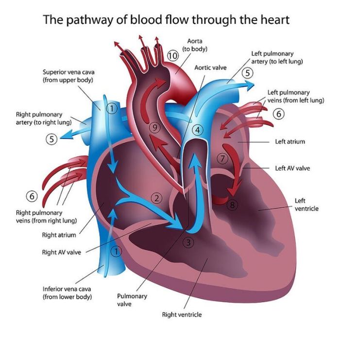
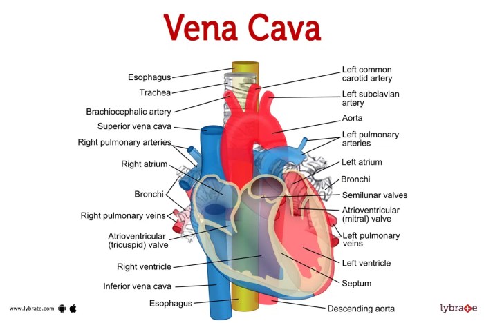
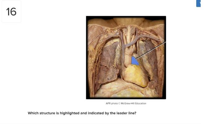
Which structure is highlighted superior vena cava – The superior vena cava is a crucial vessel in the circulatory system, responsible for transporting deoxygenated blood from the upper body to the heart. Its anatomy, function, and clinical significance are explored in this comprehensive analysis.
The superior vena cava originates from the confluence of the left and right brachiocephalic veins and descends through the mediastinum to enter the right atrium of the heart. It receives blood from the head, neck, upper limbs, and thoracic cavity.
Anatomy of Superior Vena Cava
The superior vena cava (SVC) is a large vein that drains deoxygenated blood from the upper body into the right atrium of the heart. It is located in the mediastinum, the space between the lungs. The SVC begins at the confluence of the right and left brachiocephalic veins, which drain blood from the head, neck, and upper limbs.
It then descends vertically through the mediastinum, lying anterior to the right bronchus and pulmonary artery. The SVC enters the right atrium of the heart through the superior caval foramen.
The SVC receives blood from a number of tributaries, including the:
- Azygos vein
- Hemiazygos vein
- Accessory hemiazygos vein
- Right and left superior intercostal veins
- Right and left internal thoracic veins
- Pericardiophrenic vein
- Mediastinal veins
The following diagram summarizes the anatomy of the superior vena cava:
[Diagram of the superior vena cava]
Function of Superior Vena Cava

The superior vena cava plays a vital role in the circulatory system by transporting deoxygenated blood from the upper body to the heart. This blood is then pumped to the lungs, where it is oxygenated and returned to the heart via the pulmonary veins.
The SVC also helps to regulate blood flow to the heart by adjusting its diameter in response to changes in blood pressure.
Clinical Significance of Superior Vena Cava: Which Structure Is Highlighted Superior Vena Cava

There are a number of disorders that can affect the superior vena cava, including:
- Superior vena cava syndrome
- Superior vena cava thrombosis
- Superior vena cava stenosis
- Superior vena cava atresia
Superior vena cava syndrome is a condition that occurs when the SVC is obstructed, causing blood to back up in the upper body. This can lead to a number of symptoms, including:
- Swelling of the face, neck, and upper limbs
- Cyanosis (bluish discoloration of the skin)
- Dyspnea (shortness of breath)
- Cough
- Hoarseness
Superior vena cava thrombosis is a condition that occurs when a blood clot forms in the SVC. This can lead to a number of symptoms, including:
- Sudden onset of swelling in the face, neck, and upper limbs
- Cyanosis
- Dyspnea
- Chest pain
- Cough
Superior vena cava stenosis is a condition that occurs when the SVC is narrowed. This can lead to a number of symptoms, including:
- Swelling of the face, neck, and upper limbs
- Cyanosis
- Dyspnea
- Fatigue
- Lightheadedness
Superior vena cava atresia is a rare condition that occurs when the SVC is absent. This can lead to a number of symptoms, including:
- Cyanosis
- Dyspnea
- Heart failure
The diagnosis of superior vena cava disorders is based on a physical examination and a variety of imaging tests, such as:
- Chest X-ray
- Computed tomography (CT) scan
- Magnetic resonance imaging (MRI) scan
The treatment of superior vena cava disorders depends on the underlying cause. Treatment options may include:
- Medication to dissolve blood clots
- Surgery to remove a blood clot or to widen the SVC
- Radiation therapy to shrink a tumor that is compressing the SVC
Comparison with Other Structures
The superior vena cava is one of two large veins that return blood to the heart. The other vein is the inferior vena cava, which drains blood from the lower body. The superior and inferior vena cavae are similar in their function, but they differ in their anatomy and clinical significance.
The following table compares the key features of the superior and inferior vena cavae:
| Feature | Superior Vena Cava | Inferior Vena Cava |
|---|---|---|
| Location | Mediastinum | Abdominal cavity |
| Tributaries | Right and left brachiocephalic veins, azygos vein, hemiazygos vein, accessory hemiazygos vein, right and left superior intercostal veins, right and left internal thoracic veins, pericardiophrenic vein, mediastinal veins | Right and left common iliac veins, right and left renal veins, right and left gonadal veins, right and left hepatic veins, right and left adrenal veins, right and left lumbar veins |
| Clinical significance | Superior vena cava syndrome, superior vena cava thrombosis, superior vena cava stenosis, superior vena cava atresia | Inferior vena cava thrombosis, inferior vena cava stenosis, inferior vena cava atresia |
Developmental Aspects of Superior Vena Cava

The superior vena cava develops from the embryonic cardinal veins. The right and left anterior cardinal veins fuse to form the right and left common cardinal veins, which then fuse to form the superior vena cava. The superior vena cava is initially located in the neck, but it descends into the mediastinum as the embryo develops.
There are a number of congenital anomalies that can affect the development of the superior vena cava. These anomalies can range from minor variations in anatomy to complete absence of the SVC. Most congenital anomalies of the SVC are asymptomatic, but some can cause serious health problems.
The following are some of the most common congenital anomalies of the superior vena cava:
- Persistent left superior vena cava
- Right-sided superior vena cava
- Double superior vena cava
- Absence of the superior vena cava
Persistent left superior vena cava is a condition in which the left anterior cardinal vein fails to regress during embryonic development. This results in a left-sided SVC that drains into the right atrium of the heart. Right-sided superior vena cava is a condition in which the right anterior cardinal vein fails to regress during embryonic development.
This results in a right-sided SVC that drains into the right atrium of the heart. Double superior vena cava is a condition in which both the right and left anterior cardinal veins persist during embryonic development. This results in two SVCs, one that drains into the right atrium and one that drains into the left atrium of the heart.
Absence of the superior vena cava is a rare condition in which the SVC is completely absent. This can lead to a number of serious health problems, including heart failure and death.
Key Questions Answered
What is the function of the superior vena cava?
The superior vena cava transports deoxygenated blood from the upper body to the heart.
What are the common disorders that affect the superior vena cava?
Superior vena cava syndrome, thrombosis, and congenital anomalies are common disorders that can affect the superior vena cava.
How is superior vena cava syndrome diagnosed?
Superior vena cava syndrome is diagnosed based on symptoms, physical examination, and imaging tests such as chest X-ray or CT scan.